Article Text

Abstract
Objectives To operationalise fairness in the adoption of medical artificial intelligence (AI) algorithms in terms of access to computational resources, the proposed approach is based on a two-dimensional (2D) convolutional neural networks (CNN), which provides a faster, cheaper and accurate-enough detection of early Alzheimer’s disease (AD) and mild cognitive impairment (MCI), without the need for use of large training data sets or costly high-performance computing (HPC) infrastructures.
Methods The standardised Alzheimer’s Disease Neuroimaging Initiative (ADNI) data sets are used for the proposed model, with additional skull stripping, using the Brain Extraction Tool V.2approach. The 2D CNN architecture is based on LeNet-5, the Leaky Rectified Linear Unit activation function and a Sigmoid function were used, and batch normalisation was added after every convolutional layer to stabilise the learning process. The model was optimised by manually tuning all its hyperparameters.
Results The model was evaluated in terms of accuracy, recall, precision and f1-score. The results demonstrate that the model predicted MCI with an accuracy of 0.735, passing the random guessing baseline of 0.521 and predicted AD with an accuracy of 0.837, passing the random guessing baseline of 0.536.
Discussion The proposed approach can assist clinicians in the early diagnosis of AD and MCI, with high-enough accuracy, based on relatively smaller data sets, and without the need of HPC infrastructures. Such an approach can alleviate disparities and operationalise fairness in the adoption of medical algorithms.
Conclusion Medical AI algorithms should not be focused solely on accuracy but should also be evaluated with respect to how they might impact disparities and operationalise fairness in their adoption.
- artificial intelligence
- neural networks, computer
- medical informatics applications
Data availability statement
Data was obtained from an open access database, which is publicly available. Not applicable.
This is an open access article distributed in accordance with the Creative Commons Attribution Non Commercial (CC BY-NC 4.0) license, which permits others to distribute, remix, adapt, build upon this work non-commercially, and license their derivative works on different terms, provided the original work is properly cited, appropriate credit is given, any changes made indicated, and the use is non-commercial. See: http://creativecommons.org/licenses/by-nc/4.0/.
Statistics from Altmetric.com
Summary box
What is already known?
Most prior studies on early Alzheimer’s disease (AD) and mild cognitive impairment (MCI) detection have used a three-dimensional (3D) convolutional neural networks (CNN) approach.
The 3D CNN approach is computationally expensive requiring high performance computing (HPC) infrastructures, and, due to the high number of parameters, it requires larger data sets for training.
A two-dimensional (2D) CNN needs less parameters, less computational power and execution time, while requires smaller data sets for training, but has not been applied to date for MCI detection.
What does this paper add?
The proposed approach based on a 2D CNN operationalises fairness in the adoption of medical artificial intelligence (AI) algorithms by providing fast, cheap and accurate-enough detection of early AD and MCI without the need for use of large data sets or costly HPC infrastructures.
The proposed approach can be extended to other diseases as well as to other cases where time is scarce, powerful computational resources are not available, and large data sets are out of reach.
Introduction
Recent studies show that artificial intelligence (AI) applications can perform on par with medical experts on MRI analysis.1 Such applications, to date, tend to oppose the accuracy of AI to the performance of clinicians. For instance, there have been more than 20 000 studies on deep learning (DL) methods for MRI analyses the last decade, which compare the performance of AI to the one of clinicians.2 Recent work suggests that future studies should focus on the comparison of performance between clinicians using AI and their performance without an AI aid.3 The recent global pandemic, however, revealed another urgent need of early disease diagnosis: the ability to make predictions based on a limited number of cases. The AI computer-aided detection (CAD) frameworks, to date, are based on large amounts of data and require high-performance computing (HPC) infrastructures. To address that lacuna, we propose a synergistic approach, in which clinicians and scientists collaborate for faster, cheaper and more accurate detection, relying on small data sets to make accurate-enough predictions. A promising frontier where AI can assist clinicians is Alzheimer’s disease (AD) since the release of promising clinical studies for a new drug have unearthed the need for its early detection. As it can take up to 20 years before patients with AD show any signs of cognitive decline, it can be challenging to diagnose AD in early stages. We, thus, motivate and implement an AI-CAD framework for the early detection of mild cognitive impairment (MCI) and AD to assist clinicians, while the approach can be extended for the diagnosis of other diseases.
AD is caused by an accumulation of β-amyloid (Aβ) plaques, and abnormal amounts of tau proteins in the brain. This results in synapse loss, where the impulse does not reach the neurons, and in loss of structure or function of neurons, including their death, causing memory impairment and other cognitive problems.4 AD has strong impact on the cognitive and physical functioning of patients, resulting in death. Recent developments in slowing AD decline have increased the relevance of its early detection,5 and MCI plays an important role in this. MCI is a syndrome where the patients have greater cognitive decline than normally expected, but it does not necessarily affect their daily lives. Although some patients with MCI remain stable or return to cognitively normal (CN), there is a 10%–15% risk per year of progression to AD.4 Before the aetiology of AD became known, its diagnosis relied on neurocognitive tests. The development of biomarkers improved AD detection. A common method to diagnose AD is hippocampus segmentation, which relates to memory function, and its small volume is an AD biomarker. For a long time, AD diagnosis was done manually by looking at the brain structure and size of the hippocampus on MRI, which requires practice and precision. Prior studies on automated methods for hippocampus segmentation have used DL approaches with promising results.6 Automated hippocampus segmentation for the diagnosis of AD and MCI, however, requires clinicians’ expertise and is sensitive to interrater and intrarater variability.6
Convolutional neural networks (CNN) can become the foundation of an AI-CAD framework for supporting clinicians in the detection of early AD and MCI, since it is a successful approach for image classification. CNN can improve the performance of image classification,7 and they are becoming increasingly popular in MRI analysis. For instance, recent studies show that CNN can work on par with specialists for classifying MRI of patients with skin cancer.1 Similar approaches with three-dimensional (3D) as well as two-dimensional (2D) CNN have also been used for AD detection with promising results. When it comes to the inner mechanics of these approaches, the classification filter of a 3D CNN slides along all the three dimensions of the input image, resulting in 3D feature maps, whereas in a 2D CNN the classification filter slides along only the height and width of the input image. Thus, the latter results in 2D feature maps, which need less parameters, computational power and execution time. Most prior studies have used 3D CNN achieving high accuracy,8 while others obtained similar results with 2D CNN.9 Although previous work on the topic has established that 3D CNN perform better for patch classifications, the results between 2D and 3D approaches for whole image labelling did not differ much.10 A 3D CNN, however, is more computationally expensive, and, due to the high number of parameters, it requires larger data sets for training.11 Concurrently, prior studies have not incorporated a 2D CNN approach for detecting MCI. A summary of prior 2D and 3D CNN applications in the literature is presented in table 1.
Performance comparison of 2D and 3D approaches in the literature
We suggest that medical algorithms should not be solely focused on accuracy but should also be evaluated with respect to how they might impact disparities and operationalise fairness in their adoption. Thus, we investigate the extent to which a 2D CNN can detect MCI and early AD.
Methods
CNN is the most common neural network (NN) architecture for image classification. Fully connected NN take multiple inputs, and hidden layers perform calculations on them, while the neurons in the network connect to each other. Neurons in CNN, however, connect only to those close to them. CNN, therefore, needs fewer parameters, which results in benefits such as small risk of overfitting, higher accuracy and faster processing time. Moreover, in CNN, there is no need to transform the input images to one dimensional, a process which can result in loss of structural information, as the CNN can learn the relationships among the pixels of input by extracting representative features with kernel convolutions4 :

where I is the input and K is the kernel; the input indices are represented by i and j , and the kernel indices are represented by m and n .
The data sets used in this study were obtained under permission from the Alzheimer’s Disease Neuroimaging Initiative (ADNI) database (adni.loni.usc.edu). The ADNI was launched in 2003 as a public–private partnership. The primary goal of ADNI has been to test whether MRI, biological markers and clinical as well as neuropsychological assessment can be combined to measure the progression of MCI and early AD. The ADNI is separated into three studies of 5 years, while the first was prolonged by 2 years under the name ADNI-GO. In total, 2517 people of ages 55–90 participated in the study. The ADNI encourages the use of their standardised data sets to ensure consistency in analysis and direct comparison of various methods among studies. We, therefore, used their two standardised data sets ‘ADNI1: complete 2 year 1.5T’ and ‘ADNI1: complete 3 year 1.5T’, which contain MRI that has passed quality control assessment.12
Our data set consists of 3312 images, distributed in 828 MRI of CN subjects, 453 MRI of patients with AD and 1203 MRI of patients with MCI. The data set was split into one with CN and AD subjects (1281 MRI), and one with CN and MCI subjects (2031 MRI). Since the participants of the ADNI study returned for more than one check-up, any patient can have up to 12 MRI, which are not identical as they are taken at different moments, and every MRI in the standardised data set was treated independently. The data set, thus, refers to 99 patients with AD, 212 patients with MCI and a control group of 165 CN subjects. We present the demographic information of the included subjects in table 2, to enable comparison with other studies.
Demographic information of subjects in the dataset
While the data sets are preprocessed, we further performed skull stripping using the Brain Extraction Tool V.2 (BET2), which is part of the NiPype library. Skull stripping locates the brain in the MRI and removes all surroundings to further remove noise from images. For optimal skull stripping results, neck slices were removed with the robustfov function. We used a fraction intensity of 0.3 as an evaluation of BET2 parameters for the ADNI data set found that this leads to best results. Due to the differences in scanners and techniques used by the ADNI over the years, the MRIs used in the data sets were of different sizes, and, therefore, had to become uniform. All the MRIs in our data set were resized to: (136, 192, 160) with the ndimage zoom function of the Scipy library, which zooms the array using spline interpolation. Resizing the MRI results in a different range of pixel values, and, therefore, to assure that the pixel values of all MRI had the same range, z-score normalisation was applied, which is defined as follows:

where
x
is the MRI data and  the
i
th normalised MRI. The data set was then split into train set, validation set and test set with a ratio of 60:20:20, respectively.
the
i
th normalised MRI. The data set was then split into train set, validation set and test set with a ratio of 60:20:20, respectively.
An NN consists of an input layer, hidden layers and an output layer. A CNN has hidden layers divided into convolution, pooling, activation and classification layers. We based our architecture on LeNet-5, which includes two convolutional layers, two pooling layers and two fully connected layers (supplementary files, table 3).
CNN architecture
We employ the Leaky Rectified Linear Unit (LReLU) as activation function for all convolutional layers, which allows for a small non-zero gradient.13 The LReLU activation function in the model, with x being the input data, is described as:

A Sigmoid activation function was applied to the dense layer, which outputs the probability of the images’ class, with 0 if healthy and 1 if not (AD or MCI). The Sigmoid activation function in the model, with x being the input data, is described as:

We optimised the model by manually tunning the hyperparameters (see table 4).
Parameter tuning on the AD dataset
The batch size was set to 16 and we used the Adam optimiser14 with a learning rate of 10-3. The model showed overfitting, which means that it includes more terms or uses more complicated approaches than necessary.15 Regularisation can control overfitting and drop-out regularisation is a commonly used approach because it is computationally inexpensive, and it prevents coadaptation among feature map units.11 In drop-out regularisation, only a fraction of the weights is learnt by the NN in each iteration. We added a drop-out layer with a value of 0.2 after each pooling layer (ie, 80% of the weights were learnt in each iteration), leading to better results on all the train, validation and test sets. To stabilise the learning process, we added batch normalisation after every convolutional layer. For each unit in a layer, the value was normalised as follows:

where a represents the activation vector of the ith layer l . Thereafter, the normalised values were scaled and shifted accordingly. After ~40 epochs, the model did not show increment in accuracy or reduction in loss, and overfitting increased, thus, we applied an early stopping at 40 epochs instead of the initial set of 50.
The CNN was built with a Jupyter Notebook using Python V.3.6.4, Tensorflow V.2.4.0 and Keras V.2.4.0. To load the data in NIfTI format, we used the Nilearn library, and we used the scikit-learn and SciPy libraries for data preprocessing. The development, testing and application of the model took place on the Google Cloud Console, where we used a storage bucket to store the data sets, and three compute engine instances to perform the skull stripping and preprocessing and to run our model independently as these steps require different computational resources. For skull stripping, we used an instance with 8 vCPUs, 52 GB RAM, and two NVIDA Tesla K80 GPUs, for preprocessing, we used an instance with 40 vCPUs and 961 GB RAM. For the CNN, we used an instance with 64 vCPUs, 416 GB RAM and four NVIDA Tesla T4 GPUs.
Results
The model was evaluated in terms of accuracy, recall, precision and f1-score. Recall provides sensitivity information on how many patients were correctly identified. Precision expresses how many of the positives that the model returns were actually positive. F1-score is the harmonic mean between precision and recall. An NN adjusts its weights to optimise the loss, which is calculated with the use of binary cross entropy loss:

where
C
represents the classes,  is the predicted probability value for class
i
and
t
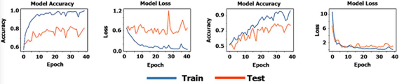
is the true probability for that class. Since the data were unevenly distributed, the accuracy baseline of random guessing was also calculated. The baseline was calculated with respect to the class distribution of the data set. First, we trained and tested our model on the AD data set. After passing the baseline of random guessing on the training data (>0.548) with an accuracy of 0.994, we applied the same model on the MCI data set. The random guessing baseline for the test data set of the AD model was 0.536 and for the test data set of the MCI model was 0.521. The overepochs performance of the model is depicted in figure 1 for AD (left) and for MCI (right).
is the predicted probability value for class
i
and
t
is the true probability for that class. Since the data were unevenly distributed, the accuracy baseline of random guessing was also calculated. The baseline was calculated with respect to the class distribution of the data set. First, we trained and tested our model on the AD data set. After passing the baseline of random guessing on the training data (>0.548) with an accuracy of 0.994, we applied the same model on the MCI data set. The random guessing baseline for the test data set of the AD model was 0.536 and for the test data set of the MCI model was 0.521. The overepochs performance of the model is depicted in figure 1 for AD (left) and for MCI (right).
Model performance for the AD and MCI datasets. AD, Alzheimer’s disease; MCI, mild cognitive impairment.
While the above graphs indicate a normal learning curve, as the performance of the model keeps increasing on the train data set, the validation performance flattens, which implies overfitting. This appears to be true mainly on the AD data set. Our model achieved accuracy of 0.837 on the AD test set. Irrespective of overfitting, the achieved test accuracy on the AD data set surpasses the random guessing baseline of 0.536. The model predicted MCI with accuracy of 0.735, passing the random guessing baseline of 0.521. Table 5 presents the performance metrics of the models on the test sets. The model performs better than chance on both sets, with a better predictive performance for the AD data set than for the MCI data set. The MCI model, however, seems to perform better on selecting relevant items (ie, recall, predicted positives relative to all positives). The MCI model shows notably less overfitting than the AD model, which might be due to the size of the data set, as the dataset used for the MCI was larger (almost double in size) than the AD one.
Performance metrics on test data
By comparing our study to previous ones in the relevant literature (see table 6), we notice a large difference in the size of the used data sets. Moreover, some of the prior studies only report the number of subjects in the used data set,8 16–18 but the number of images can differ from these since one subject can have up to 12 images in these data sets. As expected, studies with larger data sets achieved higher accuracy. Furthermore, some of the studies with a 2D approach treated the slices independently,9 19 20 thereby enlarging the size of their data set, however, the MRI was not treated as a whole.
Comparison of data and accuracy with previous studies
Discussion
While AI-CAD frameworks have been thoroughly studied, they have not been proposed as a tool for assisting clinicians. Furthermore, while the literature on AI-CAD frameworks is mostly approached from a computer science perspective, clinicians have been shown to lack trust in them.2 3 21 Our work addresses that lacuna by providing a synergistic approach between clinicians and scientists. We contribute to the line of research on using CNN for AD and MCI detection, by applying a 2D approach. Our model predicts AD better than chance by 0.301 and MCI by 0.214. As expected, the model performed worse on detecting MCI than AD. The learning process on the MCI data set, however, was much cleaner than the process on the AD data set. This might be due to the size of the data set, which can have a large impact on the process and outcomes of the model. The proposed AI-CAD framework, thus, performs better than chance for AD as well as for MCI and could assist clinicians in the early detection of AD and MCI.
We suggest that medical algorithms should not be focused solely on accuracy but should also be evaluated with respect to how they might impact disparities and operationalise fairness in terms of computational resources, when it comes to their adoption. Our framework can be further extended to other diseases, and to cases where time is scarce, computational resources are not available, and large data sets are out of reach. Finally, our work is in line with the broader Information Systems research agenda,22 on the adoption of responsible medical AI algorithms,23 and the stewardship of sensitive personal data.24 Therefore, our work can give rise to new avenues for interdisciplinary research and can become the bedrock for novel methodological advances as well as ground-breaking empirical findings on the broader topic.
Conclusion
Prior studies have used CNN to diagnose MCI and early AD, most of which applied 3D approached. The 3D CNN, however, have drawbacks that relate to needs for HPC infrastructures. Other studies have focused on detecting AD with a 2D CNN, achieving similar results as the 3D approach. Despite the relevance of detecting MCI, prior studies did not investigate how these methods perform on detecting MCI. Our main goal was to determine whether a 2D CNN can be used to diagnose AD and MCI. Our work resulted in an AI-CAD framework that can assist clinicians in the early detection of MCI and AD with high-enough accuracy, based on a relatively small data set, and without the need of HPC infrastructures. Our work has limitations that need to be acknowledged. First, an important preprogressing step is image resizing. We used Scipy ndimage, which distorts the image and could have a negative effect on the learning process. A better solution for resizing images is needed but to the best of our knowledge is not available. Second, the ADNI data sets consist of more images than participants. If subjects appear in both data sets, the model could learn subject-specific features, but the impact on model performance is unknown, as most physical features are removed during skull stripping. Third, the AD model appears to be overfitting, which is a common problem in DL models. To further optimise our model, the overfitting problem needs to be addressed by future research. Future research should also replicate the existing 3D CNN approaches and compare their execution time with the 2D CNN one of our models on the same computational infrastructure. Such a comparison will further illustrate the merits of our approach. Finally, future research should also evaluate the performance of clinicians using our framework and their performance without an AI aid.
Data availability statement
Data was obtained from an open access database, which is publicly available. Not applicable.
Ethics statements
Patient consent for publication
Ethics approval
Durham University Business School, DUBS-2021-10-31T21:05:04-xggj42.
Footnotes
Twitter @angelopoulos
Contributors LMH conceived the idea for the project and SA oversaw its overall direction and planning. Both LMH and SA worked on the acquisition of the data for analysis. LMH designed the computational framework and analysed the data. SA worked out the technical details for analysis and helped in the interpretation of the results. LMH is the guarantor of the study.
Funding The authors have not declared a specific grant for this research from any funding agency in the public, commercial or not-for-profit sectors.
Competing interests None declared.
Provenance and peer review Not commissioned; externally peer reviewed.